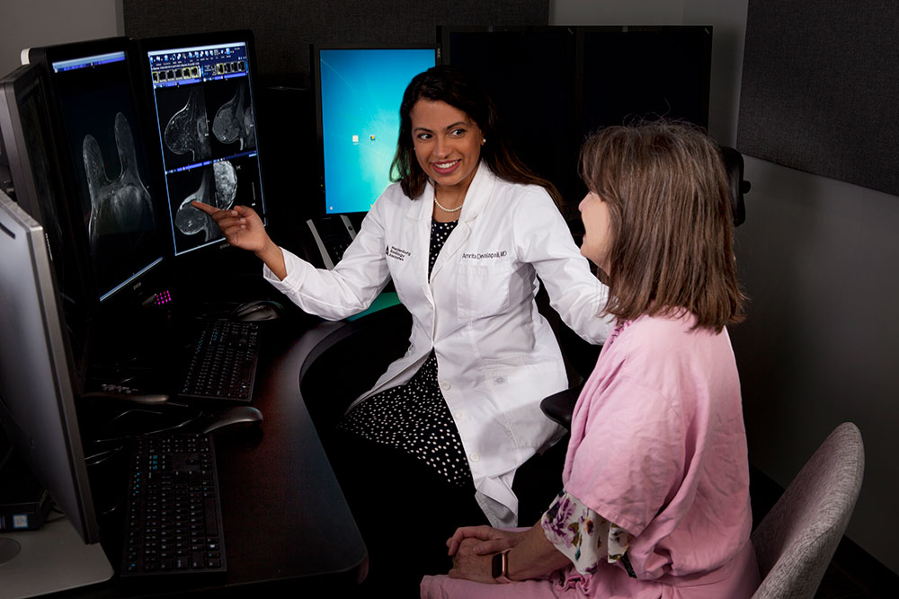
Our Breast Imaging radiologists have specialized training in detecting and treating breast disease. Having trained at several of the most advanced institutions around the country, they can bring cutting-edge tools and techniques to breast imaging centers throughout North Carolina.

We care for our patients for a lifetime. We see all of our patients for yearly mammograms and sometimes more frequently when needed. We have made our process – from screening to diagnosis – as efficient as possible so that our patients spend as little time as possible at our centers and as much time as they can enjoying life.
We continue to develop new ways to provide convenient imaging services by the radiologists you trust. We offer various breast imaging techniques at each location, including 3D mammograms and MRI. We are the right choice for women looking to get the most out of their breast imaging experience.
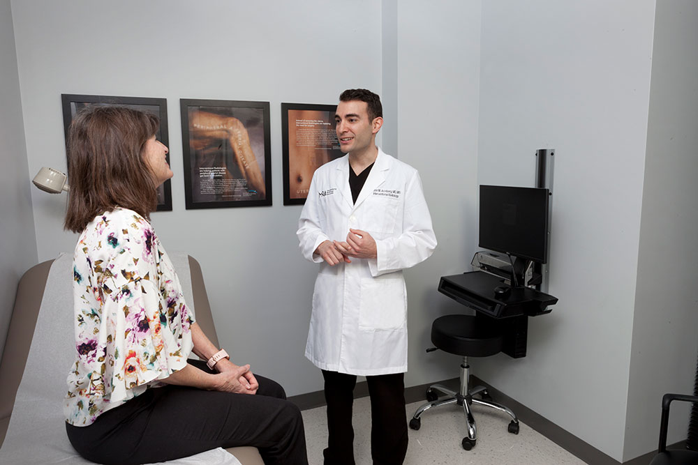
Our Breast Imaging radiologists have specialized training in detecting and treating breast disease. Having trained at several of the most advanced institutions around the country, they can bring cutting-edge tools and techniques to breast imaging centers throughout North Carolina.

We care for our patients for a lifetime. We see all of our patients for yearly mammograms and sometimes more frequently when needed. We have made our process – from screening to diagnosis – as efficient as possible so that our patients spend as little time as possible at our centers and as much time as they can enjoying life.

We continue to develop new ways to provide convenient imaging services by the radiologists you trust. We offer various breast imaging techniques at each location, including 3D mammograms and MRI. We are the right choice for women looking to get the most out of their breast imaging experience.