Diagnostic Imaging

A vital component of modern healthcare allows physicians to evaluate the inside of the body without surgery.

Diagnostic radiologists use medical imaging modalities like CT, MRI, Ultrasound, X-ray, and Fluoroscopy to diagnose and treat various conditions and diseases throughout the body. All radiologists are trained in general diagnostic radiology, and many have additional training to provide sub-specialized interpretation on any part of the body. Subspecialties of diagnostic radiology include body imaging, breast imaging, musculoskeletal radiology, and neuroradiology.

Also known as radiography, X-ray is the oldest and most commonly used type of medical imaging. X-ray uses a minimal dose of radiation to produce images of the body’s internal structures. X-rays of specific body regions help radiologists diagnose the presence or absence of disease, fractures in bones, look for foreign objects, check the positioning of lines and tubes, or other structural abnormalities. A fluoroscopy exam is a form of imaging that obtains real-time and continuous X-rays. 

Computed tomography (CT) or computed axial tomography (CAT) is a fast, non-invasive medical imaging test that uses advanced x-ray equipment to generate cross-sectional images of the body. A CT scan produces detailed images of internal organs, bones, soft tissue, and blood vessels. CT is also used to help plan and guide interventional or therapeutic procedures and monitor the effectiveness of therapy, particularly in cancer patients. Radiologists customize CT exams to answer a particular clinical question on every area of the body.

Magnetic resonance imaging (MRI) is an advanced technology that uses a powerful magnetic field to create detailed images of the body’s internal structures. It is non-invasive and does not use radiation to create images. Radiologists customize MRI exams to answer a particular clinical question on many body areas. In some cases, MRI produces more detailed images and better characterize disease than other imaging methods. MRI is also used to guide interventional procedures.

This imaging modality uses minimal amounts of radioactive material to create images of the body’s internal structures. A special camera called a Gamma camera and a computer create images that are then analyzed and interpreted by a highly trained radiologist. These tests can help diagnose various health conditions and detect disease in very early stages. Nuclear medicine is also helpful for therapeutic purposes. Services we provide include PET/CT, cardiac nuclear medicine, thyroid uptake scan, thyroid cancer treatment, bone scan, and much more.

It is also known as dual-energy x-ray absorptiometry (DEXA scan). DEXA is a simple, non-invasive test that uses a low dose x-ray on the lower spine and hips to measure bone loss and mineral density. It is the most accurate method for diagnosing osteoporosis and can help track the effects of treatment of osteoporosis. DEXA can also accurately estimate the risk of developing fractures.

Smarter Imaging Decisions

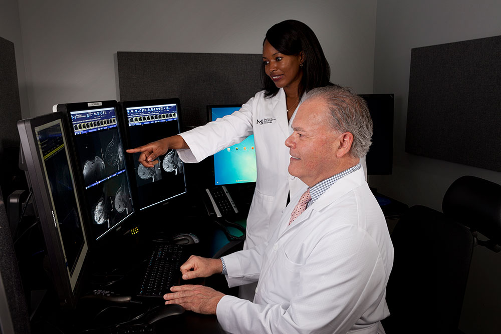
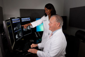
Using the latest research in healthcare imaging, we work with our patients and their doctors to make the best possible medical decisions. More information means a better understanding of the patient’s condition and more personalized treatment options.

Patient-Centered Care

With locations all over Charlotte and North Carolina, patients can expect quick and easy access to the medical imaging exam or procedure they need. Our subspecialized radiologists are on-site to intervene when a patient needs critical care, treat vein and vascular conditions, and provide expert breast imaging.

Advanced Imaging Technology

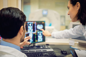
We provide comfortable, noninvasive imaging and the best image quality with the latest technology. We can diagnose a heart vessel blockage using CT, better visualize challenging neurological cases through MRI, and use image guides to provide targeted cancer treatment injections.
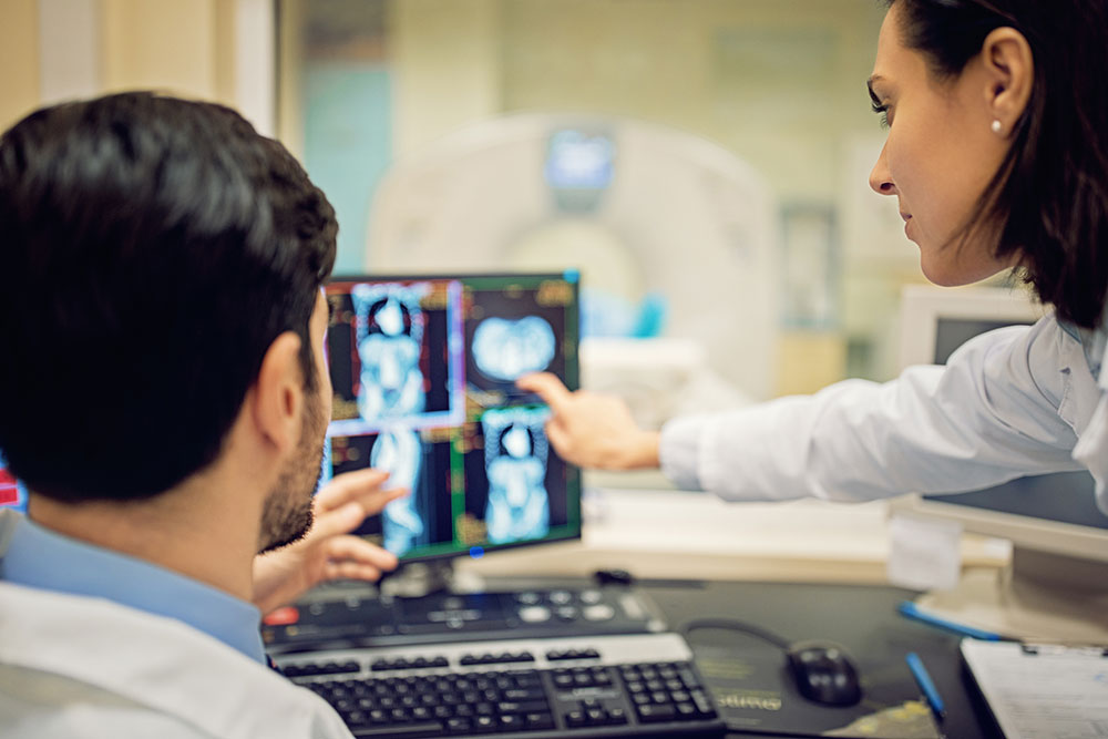